Figure 1.
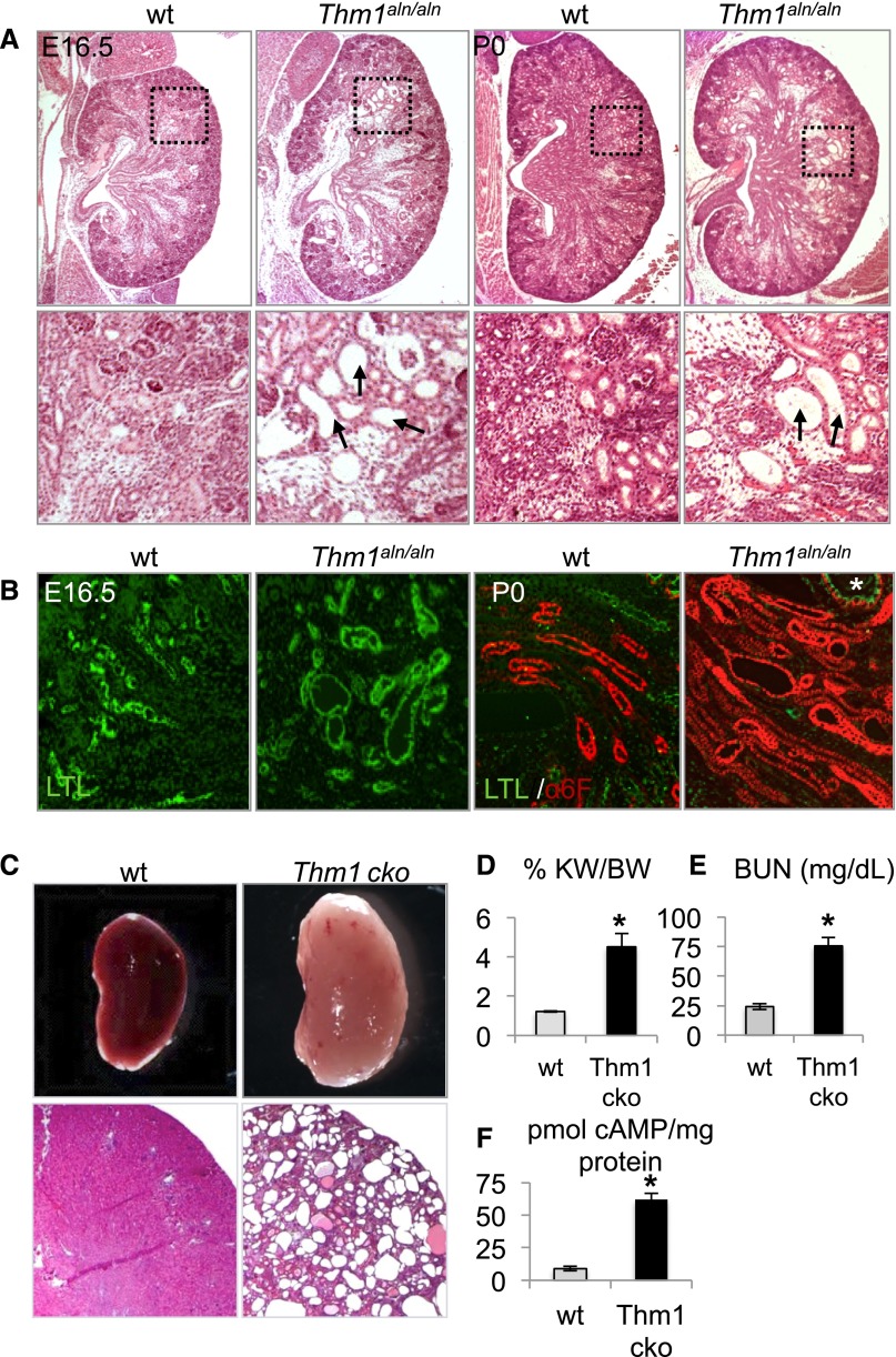
Loss of THM1 causes renal cysts. (A) Hematoxylin and eosin images of E16.5 and P0 wt and Thm1aln/aln kidneys. Black dotted squares on first-row panels are shown at higher magnification than in the second-row panels. Arrows point to dilated tubules in Thm1aln/aln kidneys. (B) Marker analysis of wt and Thm1aln/aln nephron segments. Kidney sections were co-immunostained for fluorescein-conjugated LTL (green) and for Na+K+ adenosine triphosphatase (red). (C) Whole-mount and histologic sections of 6-week-old wt and Thm1 cko kidneys. (D) %KW/BW, (E) BUN levels, and (F) cAMP levels of 6-week-old wt and Thm1 cko kidneys. Bars represent mean±SEM of 4 wt mice and 3 Thm1 cko mice. *P<0.05.

