Figure 2.
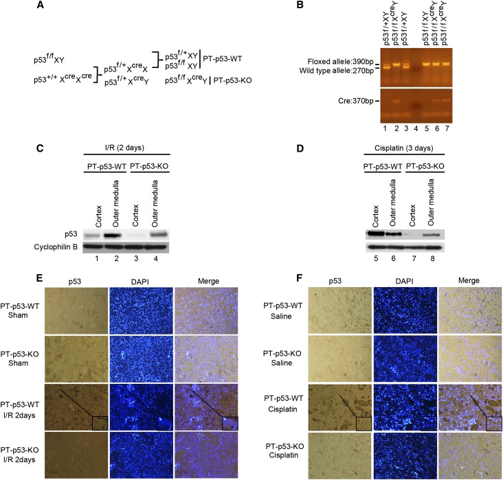
Creation and characterization of the PT-p53-KO mouse model. (A) Breeding protocol for generating PT-p53-KO mice. After confirming genotype, male littermate mice at 8–10 weeks of age were used for experiments. (B) Representative gel images of PCR-based genotyping. Genomic DNA was extracted from tail biopsy and amplified to detect wild-type and floxed alleles of p53 and PEPCK-Cre allele as indicated. (C and D) Kidney cortex and outer medulla were collected from PT-p53-KO and PT-p53-WT littermate mice after I/R injury or cisplatin injection for immunoblot analysis of p53 and cyclophilin B. (E and F) Immunohistochemical staining of p53 in kidney cortical tissues of wild-type and PT-p53-KO mice after I/R or cisplatin injury. The selected areas are shown as insets at a higher magnification. Original magnification, ×200. DAPI, 4′,6-diamidino-2-phenylindole; I/R, ischemia/reperfusion.

