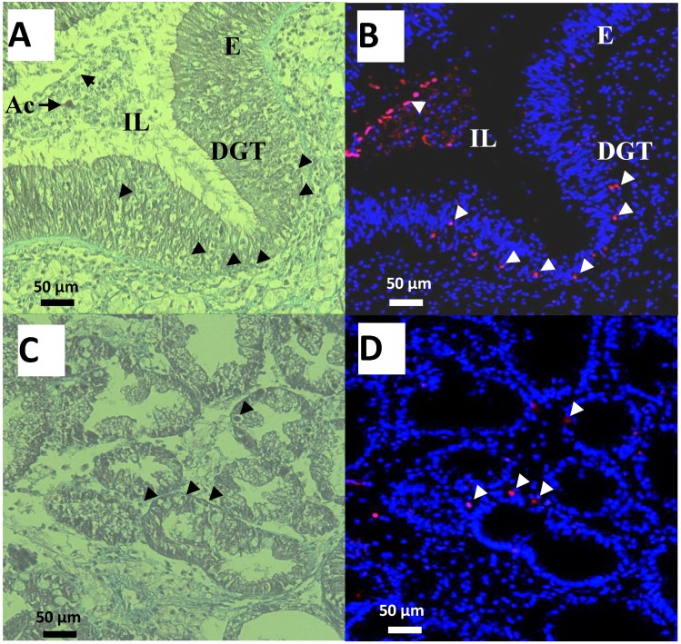
Figure 4.
Observations of cells in the digestive gland tubules (DGT), intestine epithelium (E) and Lumen (IL) of oysters exposed for 48 h to Alexandrium catenella (Ac). (A,C) Trichrome de Masson staining (B,D) Terminal deoxynucleotidyl TransferaseTetra MethylRhodamine Nick End Labeling (TTMRNEL) staining (nuclei are stained in blue, nuclei of apoptotic cells in red (Δ)).

