Figure 5.

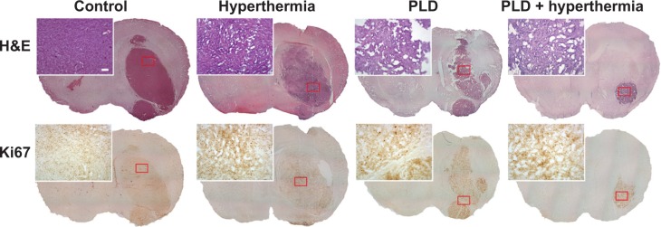
Histological (H&E) and immunohistochemical (Ki67) staining performed in tumor regions following the different treatments.
Notes: Mice were implanted with 4T1-luc2 tumor cells with treatment performed on day 6. The mice were sacrificed on day 11. Tumor slices were then obtained for staining. Tumors treated with PLD + hyperthermia had smaller tumors than the control group based on H&E staining (upper panels). Ki67 expression was associated with cell proliferation. Mild Ki67 expression was found in the tumor area of the PLD + hyperthermia-treated group. Scale bars =100 μm and 1 mm, respectively.
Abbreviations: PLD, pegylated liposomal doxorubicin; H&E, hematoxylin and eosin.
