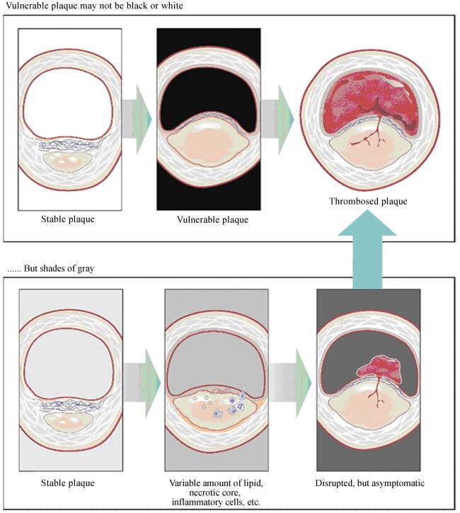
Figure 3. Vulnerable plaque.
In the upper panel, the middle figure shows a presumed vulnerable plaque, a thin capped atheroma with a large/necrotic core, and a thin fibrous cap infiltrated by inflammatory cells, which is thought to be the immediate precursor of symptomatic thrombosed plaque (upper right). However, as shown in the lower panel, a “vulnerable plaque” might not be an easy diagnosis to make with one or more invasive/noninvasive techniques. The true precursor to a symptomatic thrombosed plaque might depend on such factors as the exact cap thickness, size of the lipid/necrotic core, inflammatory cell volume, thrombogenicity of the blood, and others. Reprint with permission from reference [35].

