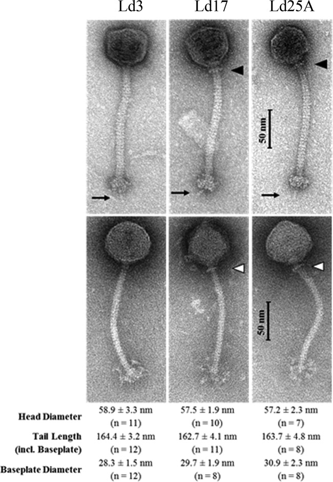
FIG 1.
Transmission electron micrographs of the L. delbrueckii subsp. bulgaricus phages stained either with uranyl acetate (UA; top three images) or with phosphotungstic acid (PTA; bottom three images). Collar-like structures of phages Ld17 and Ld25A, respectively, are indicated by triangles. The central tail fiber at the bottom of the baseplates (UA staining) is indicated by arrows. Flexible globular or fluffy appendices of baseplate structures were visualized by PTA staining.

