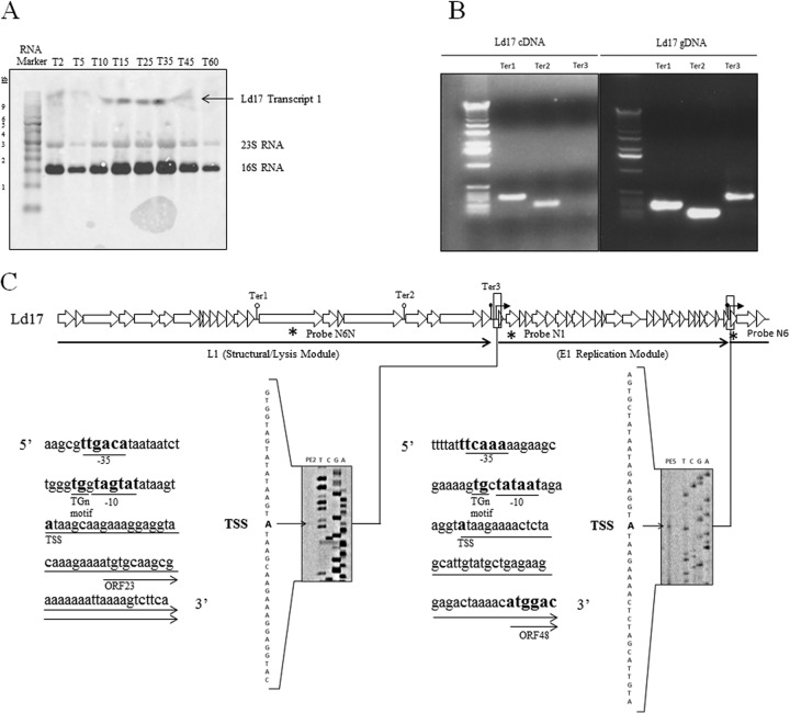
FIG 4.
Northern hybridization, RT-PCR, and primer extension analyses. (A) Northern hybridization of probe N1 to total RNA of Ld17 after infection with Ld17 at an MOI of 0.1. The time (in minutes) postinfection is indicated by the numbers along the top, with the sizes of the marker transcripts of the BrightStar Biotinylated Millenium Marker (Ambion) indicated down the left hand side. Bands due to nonspecific hybridization to rRNA are marked. (B) RT-PCR products for Ter1, Ter2, and Ter3. The left panel represents products amplified from cDNA; the right panel shows the products amplified from the phage genomic DNA control. (C) Primer extensions at the 5′ ends of transcripts E1 and L1. Transcription start sites are marked in boldface, along with the −35 and −10 promoter recognition sequences.

