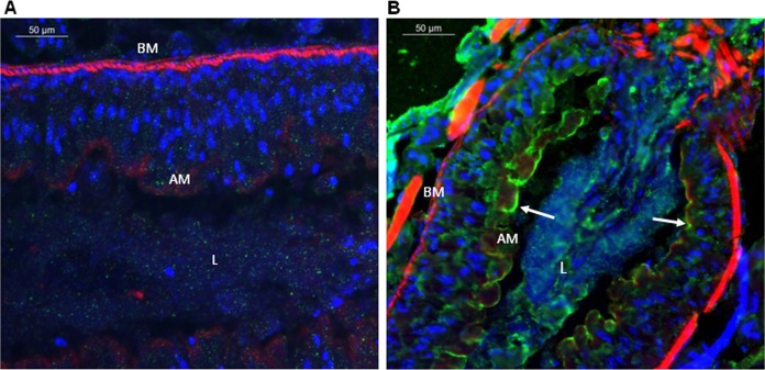
FIG 1.
Immunolocalization of Vip3Aa in midgut tissue sections (10 μm) after in vivo ingestion by S. frugiperda larvae. Binding of Vip3Aa was revealed by Alexa Fluor-conjugated secondary antibody (green) using fluorescence microscopy. Nuclei were stained with DAPI (blue), and the apical and the basal membranes were stained with phalloidin (red). Magnification × 400. (A) Larvae not exposed to Vip3Aa; (B) larvae that ingested Vip3Aa. BM, basal membrane; AM, apical membrane; L, gut lumen. White arrows show the Vip3Aa protein bound to the midgut apical membrane.

