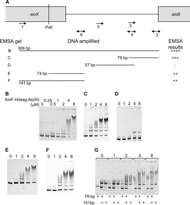
FIG 4.
Analysis of AioF-His tag-As(III) binding to different regions of the aioX-aioB intergenic region by gel mobility shift assays. (A) Schematic representation of the DNA fragments analyzed. DNA substrates for band shift assays were produced by PCR amplification using a 5′ Cy5-labeled reverse oligonucleotide (Eurogentec) (see Table S1 in the supplemental material), except that the 74-bp fragment was generated by PstI digestion of the 141-bp amplicon. The locations of the oligonucleotides used to amplify the different DNA fragments are indicated as arrows. The cyanide 5 is indicated as a black diamond. The size of the amplicon, as well as the EMSA gel reference and the EMSA results, is given. The sizes of the different DNA fragments incubated with AioF-His tag-As(III) (0, 0.25, 0.5, 1, 2, 4, or 8 μM) and tested by EMSAs were as follows: 306 bp (B), 79 bp (C), 57 bp (D), 74 bp (E), 141 bp (F), and both 79 and 141 bp (G).

