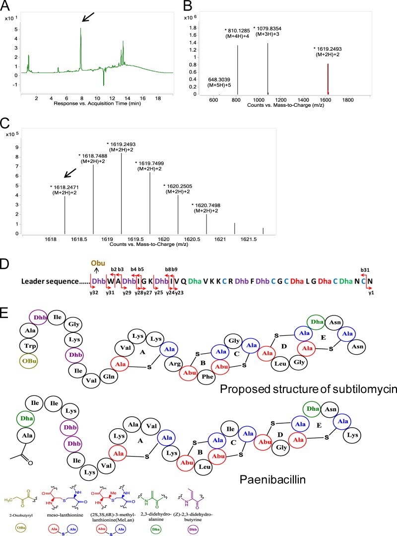
FIG 1.
Detection of the production of subtilomycin by liquid chromatography-electrospray ionization-mass spectrometry (LC-ESI-MS). (A) Chromatography profile of active ammonium sulfate crude extracts of the Bacillus subtilis BSn5 culture supernatant. The target compound is indicated by an arrow. (B) ESI-MS of the compound indicated by the arrow in panel A. (C) Magnification of the doubly charged ions [M + 2H]2+ in panel B; the monoisotopic mass peak is indicated by an arrow. (D) Fragmental ions obtained from target tandem mass spectrometry of the peptide with a mass of 3,234.48 Da. Ions y and b indicate the fragmented peptides extended from the carboxyl terminus and amino terminus, respectively. (E) Predicted structure of subtilomycin and established structure of paenibacillin (15). The predicted structure motifs are shown in different colors. Obu refers to 2-oxobutyryl.

