
FIG 6.
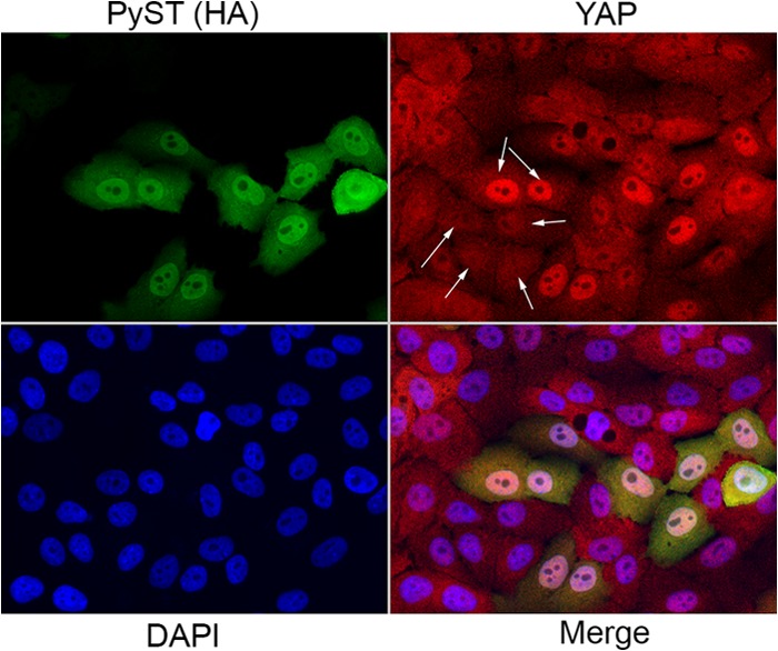
PyST promotes nuclear localization of YAP. (A) YAP localization in the presence of PyST was analyzed by immunofluorescence. MCF10a cells were infected with retroviruses expressing PyST. Fixed cells were stained with YAP antibody (shown in red; the top two arrows show highly concentrated YAP in the nucleus when PyST is expressed, and the bottom four arrows indicate that YAP is dispersed throughout the cell in cells that did not express PyST). PyST expression was detected with an HA tag antibody (shown in green). DAPI was used to stain the nucleus (shown in blue). A panel showing all three channels (Merge) is also shown.