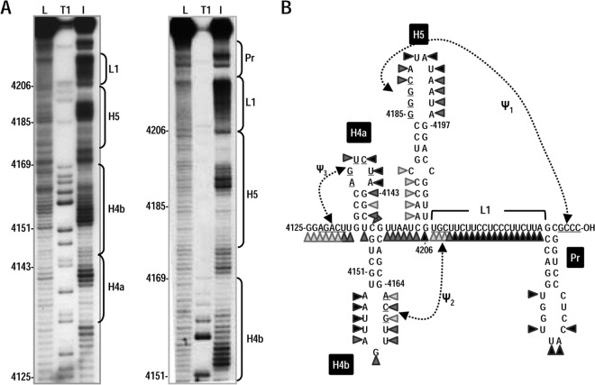
FIG 2.
In-line probing of the 3′ end of PEMV. (A) Susceptibility of residues at the 3′ end of PEMV to in-line cleavage. The 170-nt 3′ terminal fragment was radiolabeled at the 5′ end and incubated at 25°C for 14 h, followed by denaturing gel electrophoresis. The locations of different putative 3′ elements are indicated to the right of each autoradiogram. L, OH− treated ladder; T1, partial RNase T1 digest to denote location of guanylates; I, in-line cleavage of the fragment. The intensity of each band is proportional to the flexibility of the residue at that location. (B) Susceptibility of residues in the putative structure of PEMV 3′ region to in-line cleavage. Darker triangles denote stronger cleavage.

