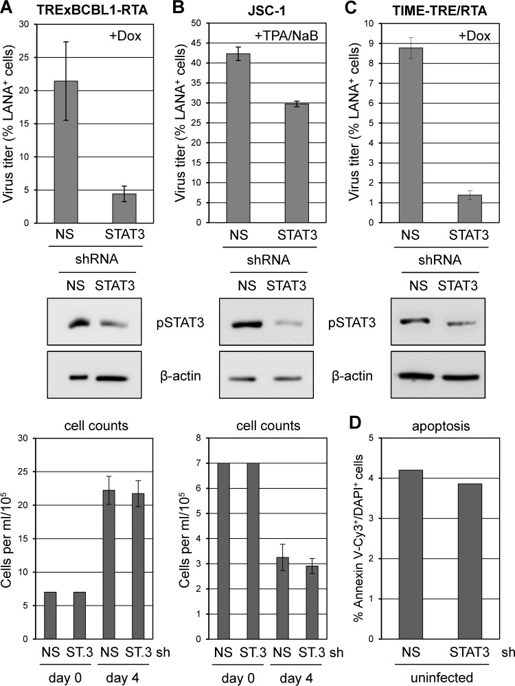
FIG 4.
Role of STAT3 in HHV-8 replication in PEL and endothelial cells. (A) TRExBCBL1-RTA cells were transduced with control (NS) and STAT3 (ST.3)-specific shRNA-encoding lentiviral vectors. After 2 days, lytic reactivation was induced with doxycycline for 4 days, and virus was then collected and concentrated from culture media for titration. Experimental procedures and data collection and calculations were as outlined in the legend to Fig. 1. Subsets of transduced cells were harvested at the time of induction and lysed for Western blot analysis to verify STAT3 depletion. Densities of viable (trypan blue-excluding) cells were normalized at the time of lytic induction (day 0) and determined at the end of the experiment (day 4) (bottom panel). (B) The same analyses were undertaken for JSC-1 cultures induced with TPA/NaB and harvested after 4 days for virus titration. A subset of cells was lysed for Western blotting at the time of induction to verify STAT3 depletion. Densities of viable cells at the start and end of the experiment are shown (bottom). (C) Doxycycline-induced TIME-TRE/RTA cultures were analyzed in the same way except that virus was harvested following 6 days of doxycycline treatment; parallel cultures were harvested at the time of induction for Western blot analysis. Cultures were not visibly affected by STAT3 depletion during the course of infection (not shown). (D) Rates of apoptosis (% annexin V-Cy3+/DAPI+ cells) in similarly transduced TIME-TRE/RTA cultures were unaffected by STAT3 depletion.

