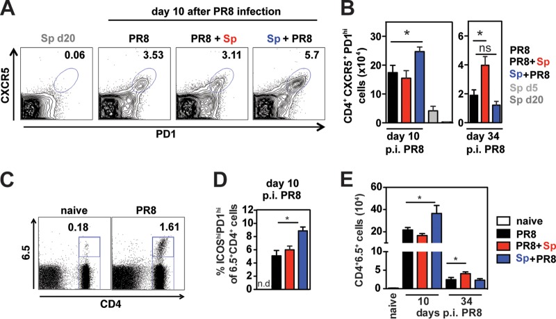
FIG 3.
Bacterial coinfection alters T follicular helper cell responses in the medLN of influenza virus-infected mice. (A) Representative FACS profiles of CXCR5 and PD1 staining on CD4+ T cells in the medLN in indicated groups of mice. (B) CXCR5+ PD1+ CD4+ TFH cells in medLN of infected and coinfected mice at indicated days after PR8 infection. Data represent means ± SEM from n = 3 to 5 mice and are the results from 2 independent experiments. Significance was determined by Student's t test. NS, nonsignificant; *, P < 0.05. (C to E) PR8-HA-specific CD4+ T cells from TS1 mice were adoptively transferred 1 day prior to PR8 infection. (C) Representative FACS profile of medLN cells from a naïve mouse and a PR8-infected mouse (10 days p.i. with PR8) stained for CD4 and the TCR-specific Ab 6.5. (D) Frequency of ICOShi PD1hi cells of CD4+ 6.5+ T cells in the medLN of infected/coinfected mice 10 days after PR8 infection (n = 3 naive mice and n = 5 to 11 infected mice/group). n.d., not determined. (E) CD4+ 6.5+ T cell numbers in the medLN of naive and infected/coinfected mice 10 days and 34 to 35 days after PR8 infection. Significance was determined by Student's t test. ns, not significant; *, P < 0.05.

