FIG 4.
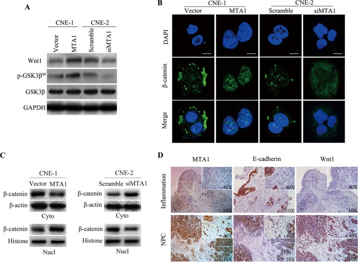
MTA1 mediates EMT, migration, and invasion by enhancing the expression of biologically active Wnt1 in NPC cell lines. (A) Representative Western blot from three independent experiments of CNE-1-vector, CNE-1-MTA1, CNE-2-scramble, and CNE-2-siMTA1 cells for the expression of Wnt1 and GSK3β phosphorylation analysis. The quantification of the Western blot signals are analyzed (data not shown). (B) Confocal image of β-catenin (green) expression in CNE-1-vector, CNE-1-MTA1, CNE-2-scramble, and CNE-2-siMTA1 cells. The scale bar depicts 10 μm. (C) CNE-1-vector, CNE-1-MTA1, CNE-2-scramble, and CNE-2-siMTA1 cells were subjected to nuclear (Nucl) and cytoplasmic (Cyto) extract isolation. The blots are representative of two independent experiments. The quantification of the Western blot signal for β-catenin was normalized to either β-actin or histone compared to untreated controls (data not shown). (D) The expression of MTA1, E-cadherin, and Wnt1 was determined by immunohistochemical analysis in inflammatory or NPC tumor biopsy specimens. The insets show images at a higher magnification. Positive staining is visible as brown spots.

