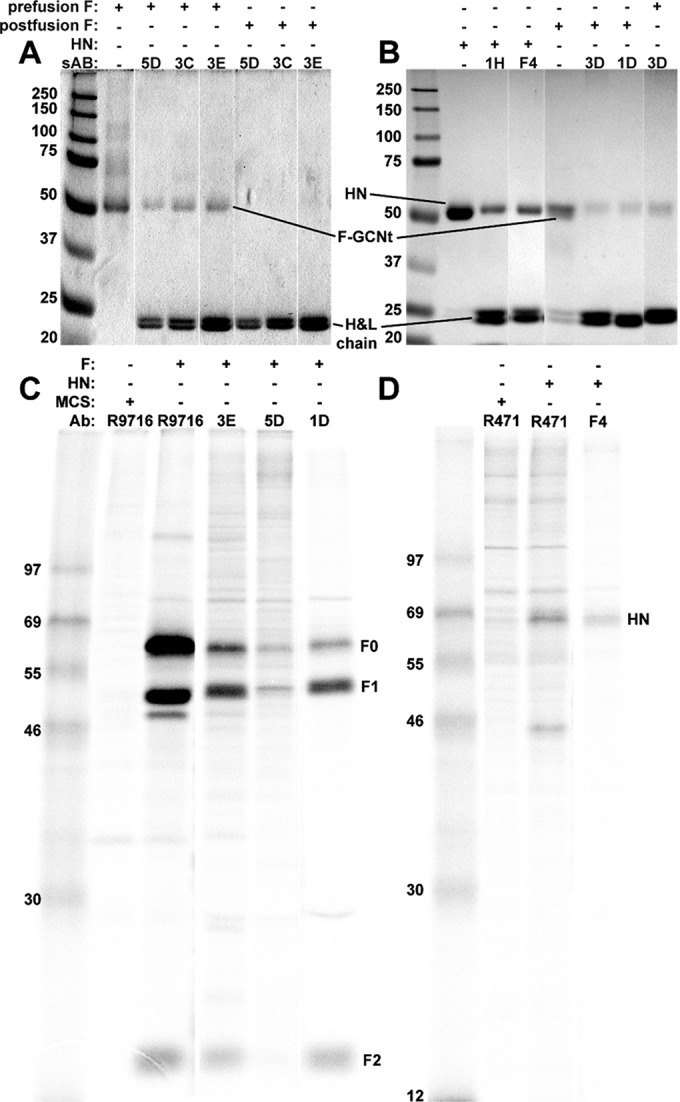
FIG 4.

Immunoprecipitation (IP) of various sAbs. (A) IP followed by reducing SDS-PAGE and Coomassie staining of the purified prefusion forms of F-GCNt using prefusion conformation-specific sAbs (third to fifth lanes). These sAbs did not immunoprecipitate F-GCNt in the postfusion conformation (sixth to eighth lanes). (B) IP followed by reducing SDS-PAGE and Coomassie staining of HN56–565 using anti-HN-specific sAbs (third and fourth lanes) and IP of the postfusion form of F-GCNt using anti-postfusion-specific sAbs (sixth and seventh lanes). 3D was able to immunoprecipitate both forms of F-GCNt (sixth and eighth lanes). (C) IP and SDS-PAGE of radiolabeled PIV5 F from transfected cells using an anti-F pAb (lane 3), anti-prefusion-specific sAbs (fourth and fifth lanes), and an anti-postfusion-specific sAb (sixth lane). (D) IP and SDS-PAGE of radiolabeled PIV5 HN from transfected cells using an anti-HN-specific pAb (third lane) and an anti-HN-specific sAb (fourth lane). H&L chain, heavy and light chains. Values to left of panels indicate molecular masses (in kilodaltons).
