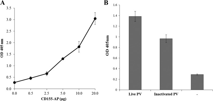
FIG 1.
Analysis of the interaction between PV and soluble hPVR-AP. (A) Live PV2 (50 D-Ag) was incubated with soluble hPVR-AP (0.5 to 20 μg) at 4°C for 120 min. Samples were then ultracentrifuged through a 30% sucrose cushion. The resulting pellets were resuspended in Tris-HCl, and the amount of bound CD155-AP was quantified by a colorimetric AP reaction. The average optical density (OD) values at 405 nm of two determinations are shown with the standard error. (B) Live and inactivated MEF-1 preparations (50 D-Ag) were incubated with soluble hPVR-AP (10 μg) and processed as described above. The results were compared to those with samples centrifuged with soluble hPVR-AP alone (column “−”). The average OD values at 405 nm of three determinations are shown as bars with standard errors.

