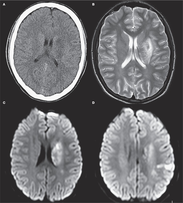
Figure 5.
A) Axial CT scan at day 1 shows a limited area of infarction in the left basal ganglia. T2 weighted (B) and diffusion-weighted (C,D) MRI at day 7: images show a limited area of infarction in the left basal ganglia and parietal cortex. A limited area of infarction was also noticed in the mesencephalon, not shown in this figure.

