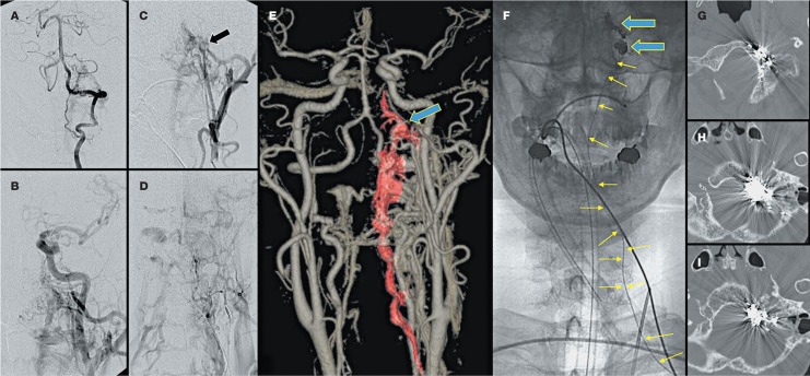
Figure 3.
Case 6. A patient suffered from vascular tinnitus for 1 year. Diagnostic angiographies of left vertebral (A), left common carotid (B), left external carotid (C) show a DAVF in the left hypoglossal canal. The drainage is mainly to the internal paraspinal venous plexus (D). On virtual reality images (E) of the CT angiogram, the fistula (arrow) and drainage are identified. The inferior petrosal sinus is not visualized in the above studies. The fistula is obliterated by paraspinal venous approach (small yellow arrows) with coil packing (large blue arrows). The follow-up CT study shows the coil mass inside the occipital condyle (G), hypoglossal canal (H), and lateral lower clivus (I).

