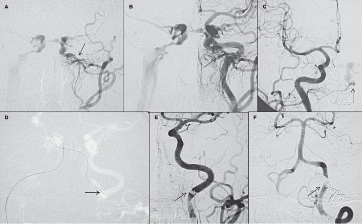
Figure 4.
Case 13. A patient had pulsatile tinnitus and left eye congestion for 1 year. Diagnostic angiography of left external (A), left common (B), and right common carotid arteries (C) show a DAVF in the left hypoglossal canal with obstruction of the lower inferior petrosal sinus. Retrograde flow shows reflux to the right cavernous sinus and right inferior petrosal sinus. The microcatheter was navigated from right inferior petrosal sinus to left inferior petrosal sinus and then the anterior condylar DAVF through the communication between both cavernous sinuses (D). Immediate post-procedure angiogram shows no residue on left common carotid (E) and left vertebral artery (F) injection.

