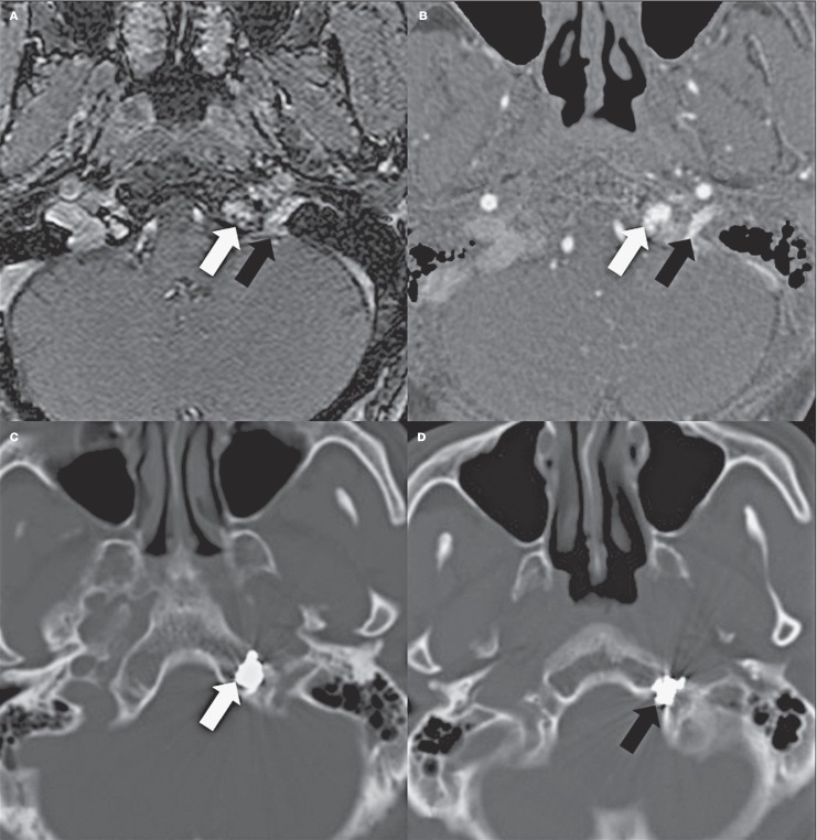
Figure 5.
Case 6. Pre-embolization time of flight MR angiography source image at the level of the hypoglossal canal showshigh flow inside the left hypoglossal canal (black arrow) and adjacent basiocciput (white arrow). It is also well depicted on the CT angiography source image (B). Post-embolization CT angiography shows a coil mass inside the basiocciput (C, white arrow) and hypoglossal canal (D, black arrow).

