FIG 1.
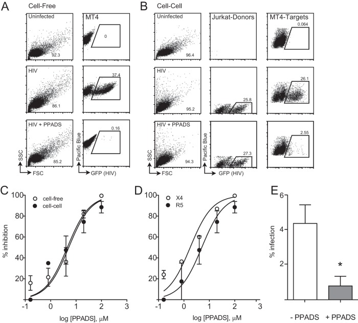
PPADS treatment results in dose-dependent inhibition of cell-to-cell and cell-free productive infection. (A) Cell-free infection is inhibited by PPADS. Representative fluorescence-activated cell sorter plots are shown for uninfected cells (top) or HIV NL-GI-infected Alexa Fluor 450-labeled MT4 cells (cell-free infection) in the absence (middle) or presence (bottom) of 100 μM PPADS. (B) Cell-cell infection is inhibited by PPADS. Representative fluorescence-activated cell sorter plots are shown for HIV NL-GI-nucleofected Jurkat (donor) cells mixed with Alexa Fluor 450-labeled MT4 (target) cells. MT4 cells mixed with uninfected Jurkat cells (uninfected, top), infected cells (middle), or infected cells in the presence of 100 μM PPADS (bottom). (C) Dose-response curves for both NL-GI cell-free and cell-cell infections by concentration of PPADS. Nonlinear regression curve fits for cell-to-cell and cell-free infection are shown. (D) Dose-response curves of NL-GI (X4) and NL-GI-RHPA (a primary R5-tropic virus cloned into the NL-GI backbone) cell-free infections, using MT4 or MT4-R5 target cells, by concentration of PPADS. Infections were conducted in the presence of serial 5-fold dilutions of PPADS from 1 mM, and samples were incubated for 36 h and then fixed and analyzed by flow cytometry. (E) PPADS inhibition of NL-GI infection of activated PBMCs after 48 h. PPADS inhibited productive infection by 82%. Results are the means ± SEMs of three independent experiments. *, P < 0.05.

