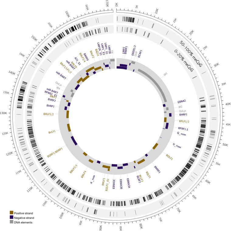
FIG 6.
Methylation of the EBV genome in stably infected NOK. The outer circle displays the EBV genome position. The inner gray circle annotates select EBV genes, with purple bars showing rightward reading open reading frames and gold bars indicating the leftward reading open reading frames. Repetitive elements and origins of DNA replication are indicated as gray bars. The light gray circles map the positions of methylated (50 to 100% meCpG) and unmethylated (0 to 20% meCpG) CpG residues.

