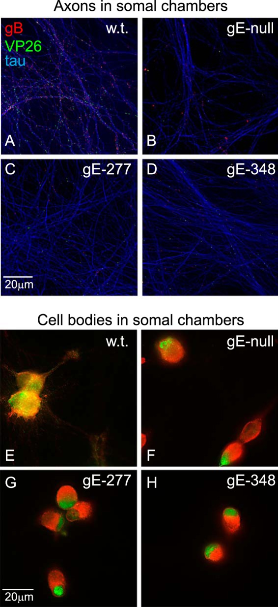
FIG 4.

Capsids and gB puncta in proximal axons and cell bodies. SCG neurons were infected as described in the legend to Fig. 2 for 18 h. The microfluidic devices were disassembled, and the neuron bodies and axons in the somal chambers were fixed with paraformaldehyde, permeabilized, and simultaneously immunostained with antibodies specific for VP26 (capsids; green), gB (red), and the microtubule-associated protein tau (purple-blue) and then with secondary fluorescent antibodies. (A to D) Images of neuronal axons, with efforts to exclude cell bodies. (E to H) Images of neuronal cell bodies and their associated proximal axons, with the tau-specific fluorescence subtracted.
