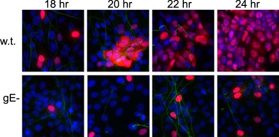
FIG 5.

Spread of wt and gE-null HSV from distal axons to adjacent epithelial cells. SCG neurons growing in microfluidic chambers were allowed to produce axons, which extended into the axonal compartment for 6 days, and then HaCaT cells were plated in the axonal compartments for 24 h. The neurons were then infected with wt HSV or HSV gE-null (using 8 PFU/cell) by adding virus to the somal compartments. After 2 to 4 h, 0.1% human gamma globulin (a source of HSV-neutralizing antibodies) was added to the axonal chambers. At the indicated time points, the devices were disassembled and cells in the axonal chambers were fixed with 4% paraformaldehyde, permeabilized with 0.1% Triton X-100, and immunostained with antibodies specific for tau (axons; green) and the HSV immediate-early protein ICP4 (HaCaT cells; red), and then with secondary fluorescent antibodies and the nuclear dye DAPI (purple-blue).
