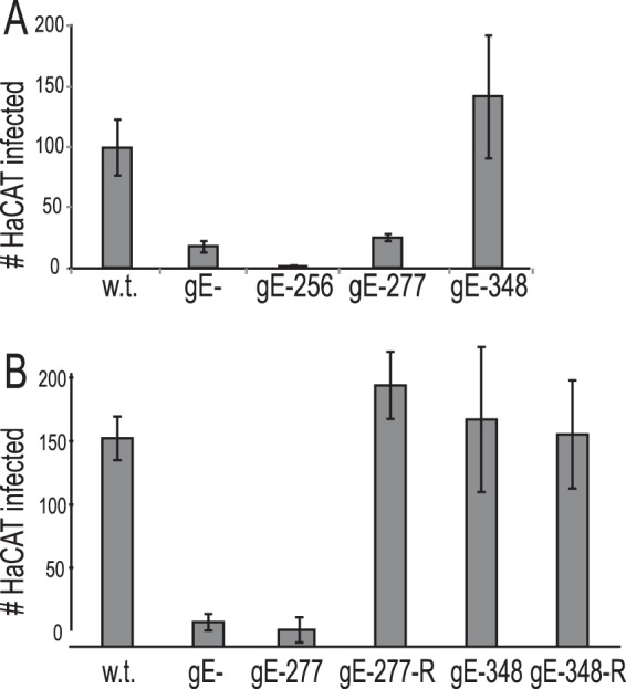
FIG 6.

Spread of F, gE−, gE-256, gE-277, and gE-348 mutants from distal axons to adjacent nonneuronal cells. SCG neurons and HaCaT cells were plated in microfluidic chambers and infected with HSV as described in the legend to Fig. 5. Cells in the axonal chambers were fixed with 4% paraformaldehyde, permeabilized with 0.1% Triton X-100, and immunostained with ICP4-specific antibodies and the nuclear dye DAPI. (A) ICP4+ HaCaT cells in 10 axonal compartments involving 3 separate wells were manually counted, and the total numbers of ICP4+ HaCaT cells/axonal compartment are shown with standard deviations. (B) Another experiment was performed as for panel A but including repaired HSV gE-277R and HSV gE348R.
