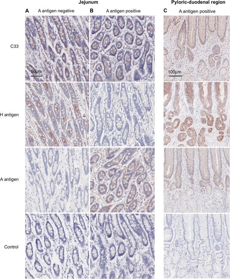
FIG 4.
Immunohistochemical analysis of CNV VLPs binding to canine intestinal tissue sections. VLPs were incubated with tissue sections overnight, and binding was detected using anti-CNV antibody and biotinylated secondary antibody. HBGA expression was determined using anti-A antigen antibody and Ulex lectin. Binding of either VLPs or antibodies/lectin is indicated by the presence of red signal. (A) Binding of CNV strain C33 to jejunal tissue from an A antigen-negative dog. (B) C33 binding to jejunal tissue from an A-positive dog. (C) Binding of C33 to tissue from the pyloric duodenal region of the intestine of an A-positive dog.

