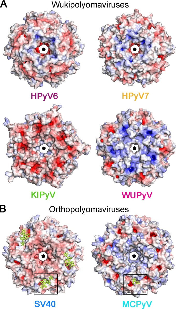
FIG 5.
Electrostatic surface potentials of VP1 pentamer from the wuki- and orthopolyomavirus genera. Overall surface representations of HPyV6, HPyV7, and KIPyV (PDB accession no. 3S7V) and WUPyV (PDB accession no. 3S7X) (A) and SV40 VP1 (PDB accession no. 3BWR) and MCPyV (PDB accession no. 4FMI) (B) pentamers are colored according to electrostatic potential (calculated using APBS tool 2.1; 46), with blue and red corresponding to +7 kT and −7 kT, respectively. Views are equivalent in panels A and B and are shown from the top—the outer surface of the virion—along the 5-fold axis of the pentamer. Carbohydrates (GM1 pentasaccharide and Neu5Ac and Gal of α2,3-sialyllactosamine) in panel B are shown in yellow stick representations, and the glycan binding site is highlighted for clarity with a box.

