FIG 1.
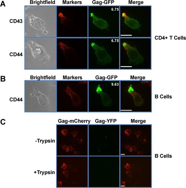
MLV Gag localizes to the uropod in polarized primary T and B lymphocytes. (A and B) Primary CD4+ T cells (A) and primary B cells (B) were infected with F-MLV Gag-GFP (green). Sixteen to 20 h after infection, cells were plated on ICAM-1-coated coverslips, immunostained for the uropod marker CD43 or CD44 (red), and examined by spinning disc confocal microscopy. Depicted images represent the merged extended-focus view of an entire Z-stack. The mean polarization indices of 10 images that quantify the accumulation of Gag-GFP at the uropod are shown at the upper right corners of the Gag-GFP images. (C) Primary B cells were infected with viruses labeled with Gag-YFP (green) and containing full-length Gag-mCherry genomes (red). Eight to 12 h postinfection, cells were treated with trypsin as indicated and monitored by spinning disc confocal microscopy. Scale bars, 10 μm.

