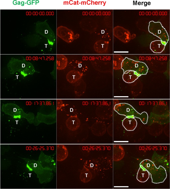
FIG 10.

An example of uropod-mediated stable virological synapses between primary B cells and T cells. Primary B cells infected with wild-type F-MLV Gag-GFP (green) as for Fig. 1B were cocultured with S49.1 T cells stably expressing receptor mCAT-1-mCherry (red) and examined by live-cell microscopy. Three image series with 8-min intervals are shown. “D” marks the donor cell and “T” marks the target cell. Scale bar, 10 μm.
