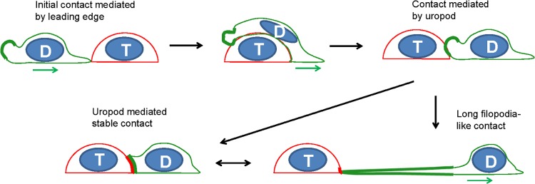
FIG 13.

Model for uropod-mediated formation of virological synapses in migrating lymphocytes. The leading edge of the migrating donor cell (green) makes an initial contact with a target cell (red), continues to migrate, and passes the target cell until a uropod-mediated contact is established. Continued migratory forces can lead to long filopodial contacts. “D” depicts the donor cell and “T” the target cell. The green and red thick lines represent the enrichment of Gag-labeled viral particles (green) and viral receptor (red), respectively. The green arrow denotes the direction of migration for donor cells.
