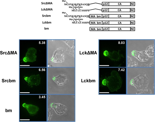
FIG 4.
Basic residues in MA are not required for MLV Gag uropod localization. Upper scheme displays tested wild-type (wt) MLV Gag and mutants of MLV Gag. Localization of indicated Gag-GFP mutants (green) within polarized primary B cells was determined as described for Fig. 1B. Scale bars, 10 μm.

