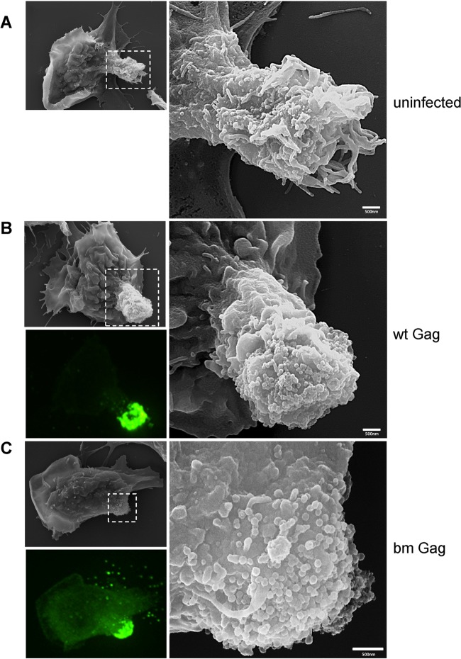
FIG 8.
SEM electrographs of polarized primary B cells reveal surface accumulation of wild-type and mutant bm Gag particles. Samples were prepared as described for TEM in the legend to Fig. 7. After fixation, virally infected Gag-GFP-labeled (green) polarized B cells were identified using fluorescence microscopy and marked using a diamond pen, and the identical cells were visualized by SEM. Images of uninfected B cells (A) and B cells infected with wild-type (B) and mutant bm Gag (C) F-MLV are displayed. The images to the right are magnifications of indicated regions. The bottom left image of each panel is the corresponding fluorescence image of MLV Gag-GFP virions.

