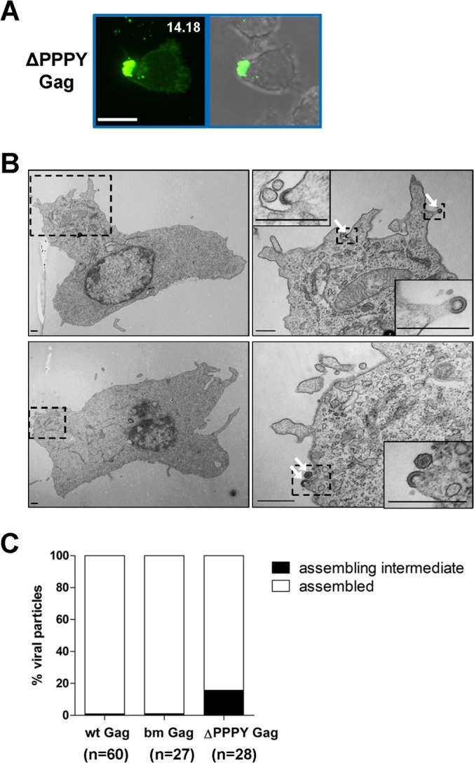
FIG 9.

Late-domain mutant virus accumulates at the uropod but fails to pinch off from the PM. (A) Primary B cells infected with F-MLV Gag-GFP ΔPPPY as described for Fig. 1B. Scale bar, 10 μm. (B) Samples were prepared as described for TEM in the legend to Fig. 7 to characterize primary B cells infected with late-domain mutant MLV (ΔΔPPPY Gag). Scale bar, 500 nm. (C) Quantification of assembly intermediates and assembled particles for wt Gag, bm Gag, and ΔPPPY Gag in TEM micrographs. Sixty, 27, and 28 polarized B cells were analyzed for wt Gag, bm Gag, and ΔPPPY Gag, respectively.
