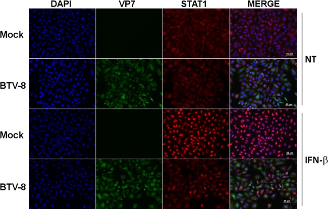
FIG 2.
BTV infection interferes with STAT1 translocation. A549 cells were mock infected or infected with 0.05 TCID50 of BTV-8/cell for 18 h and left untreated (NT) or treated with IFN-β (1,000 IU/ml) for 30 min. Cells were then washed, fixed, and stained with primary antibodies specific for VP7 and STAT1, followed by fluorescent dye-conjugated secondary antibodies. Intracellular localization of DAPI-stained nuclei (blue), VP7 (green), and STAT1 (red) was visualized by immunofluorescence microscopy (magnification, ×20). Scale bar, 50 μm.

