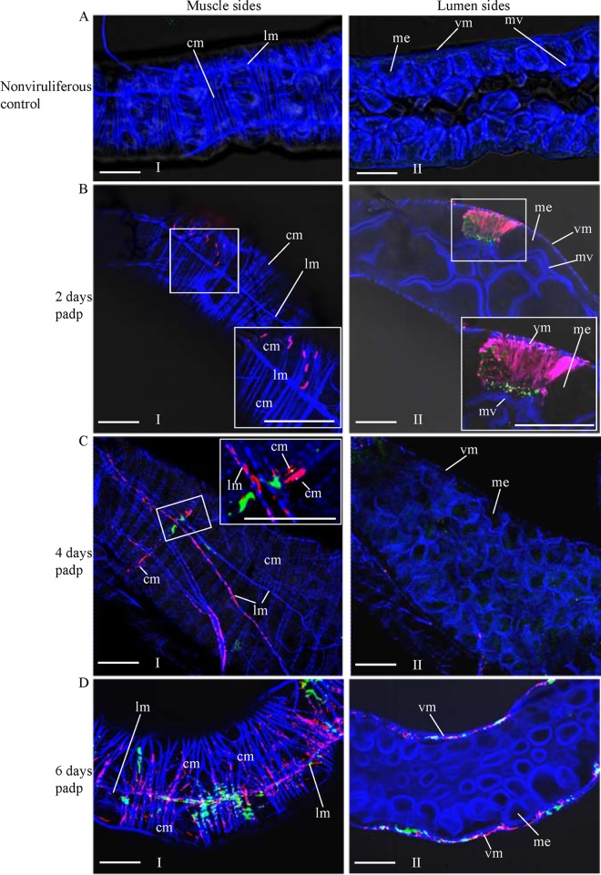
FIG 5.
Extension of P7-1 tubules from the initially infected midgut epithelium toward visceral muscle tissues in viruliferous WBPHs. The internal organs of nonviruliferous WBPHs (A) and viruliferous WBPHs at 2 days (B), 4 days (C), or 6 days (D) padp were immunolabeled with P9-1–FITC (green), P7-1–rhodamine (red), and the actin dye phalloidin-Alexa Fluor 647 carboxylic acid (blue) and then examined by confocal microscopy. (Panels I and II) the muscle (I) and lumen (II) sides of the midgut. (Insets) Enlarged images of the boxed areas. Images are representative of those from more than 4 experiments. me, midgut epithelium; mv, microvilli; vm, visceral muscle; cm, circular muscle; lm, longitudinal muscle. Bars, 30 μm.

