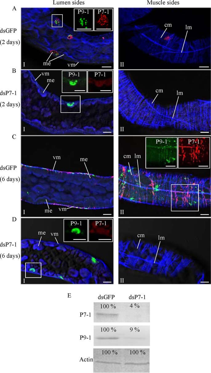
FIG 8.
Microinjection of dsP7-1 suppressed viral spread in vivo in WBPHs. The nymphs of WBPHs were microinjected with dsGFP (A, C) or dsP7-1 (B, D). At 2 days padp (A, B) or 6 days padp (C, D), the internal organs of WBPHs receiving dsGFP or dsP7-1 were immunolabeled with P9-1–FITC (green), P7-1–rhodamine (red), and the actin dye phalloidin-Alexa Fluor 647 carboxylic acid (blue) and then examined by confocal microscopy. (Panels I and II) The lumen (I) and muscle (II) sides of the midgut. (Insets) Green fluorescence (P9-1 antigens) and red fluorescence (P7-1 antigens) of the merged images in the boxed areas. Images are representative of those from more than 4 experiments. me, midgut epithelium; vm, visceral muscle; cm, circular muscle; lm, longitudinal muscle. Bars, 15 μm. (E) Detection of viral proteins P7-1 and P9-1 in WBPHs by Western blotting assay with P7-1-specific or P9-1-specific IgGs. Insect actin was detected with actin-specific IgG as a control. The protein accumulation level in WBPHs that received dsGFP was taken to be 100%.

