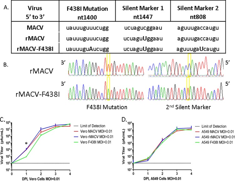
FIG 2.
Rescue and in vitro analysis of rMACV-F438I. (A) The mutation to generate the F438I amino acid change was accomplished through target PCR mutagenesis by changing the adenosine to a uracil at nt 1400. The addition of a silent mutation at nt 1407 distinguishes all rMACVs from the wild-type virus. The addition of the second marker distinguishes rMACV-F438I from rMACV. Differences between sequences are denoted by a capital letter. (B) Chromatography sequence analysis of viral cDNA confirms the presence of the F438I mutation and both silent markers. (C) TCSs from cells infected at an MOI of 0.01 with MACV, rMACV, or rMACV-F438I in triplicate (n = 3) were plaque analyzed to calculate viral titers from 0 to 4 dpi. TCSs collected from IFN-incompetent Vero cells showed similar growth of all three viruses. By 3 dpi, all three viruses reached comparable viral peaks, which were maintained at 4 dpi. (D) TCS collected from IFN-competent A549 cells had no significant difference in viral titers between all three viruses (P > 0.05, two-way ANOVA, n = 3).

