
Figure 5.
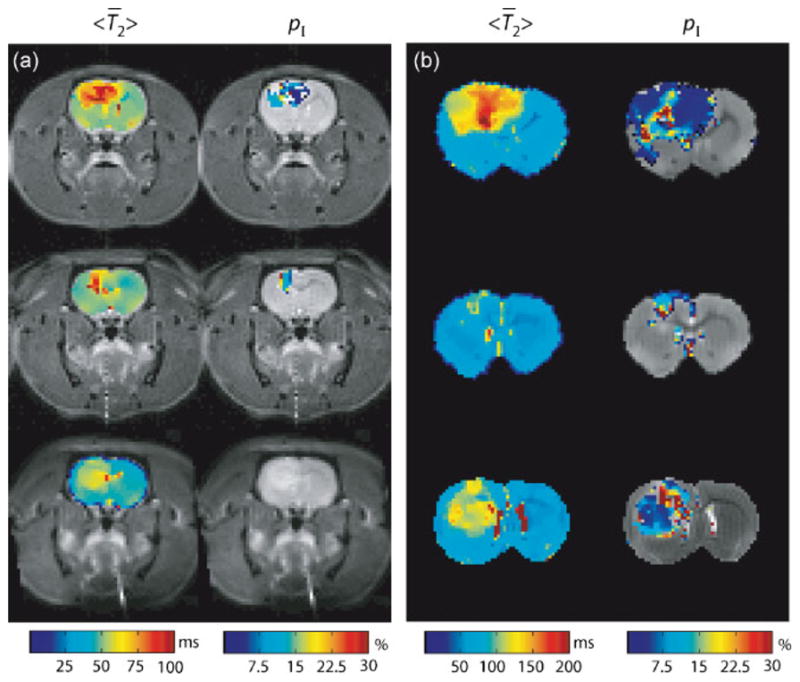
(a) In vivo <T̄2> and pI maps overlaid onto anatomical images (Rats #9–11). (b) Corresponding ex vivo results from the same three rats. For pI, only voxels whose T2 spectra were deemed admissible — defined as spectra that exhibited two components in the range 8–200 ms or 15–300 ms for in vivo and ex vivo acquisition, respectively — are shown. Additional contributions from white matter were also removed (see Methods).