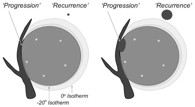
Figure 1.

Graphical representation of ablation nomenclature (25) for local tumor recurrences: “progression” versus “recurrence.” Left image shows a 4- to 5-cm tumor with five cryoprobes (small black circles) placed within 1 cm of the tumor margin and less than 2 cm apart (10). A small portion of residual unablated tumor grows as local tumor progression in the right image. A subclinical focus or “satellite” may lie less than 1 cm beyond the visible ice margin and grow as a mass adjacent to the involuting PCA zone but is still considered a local tumor recurrence.
