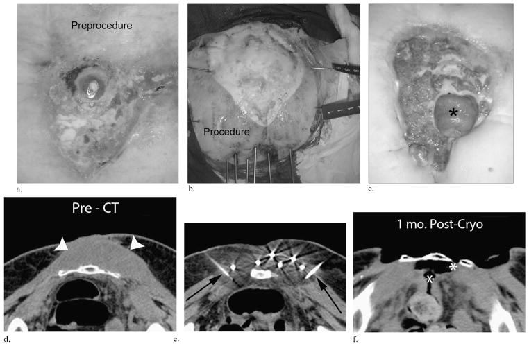
Figure 2.
(a–c) Clinical images over time in a patient with a superficial melanosarcoma invading the sacrum that had repeatedly recurred after multiple surgeries. The initial open tumor wound (a), the grossly visible ice extension beyond all tumor margins at the completion of the first freeze with six inferior cryoprobes and lateral saline solution injection needles (b), and the 3-month follow-up (c) after resection of the lower portion of the sacrum finally show a herniated underlying loop of small bowel (black asterisk) and a new satellite tumor recurrence along its inferior margin. (d–f) Companion CT images: pre-CT image through the upper mid-sacrum (d) at the maximum tumor extent (arrowheads). During PCA (e), ice rapidly extended posteriorly into the rectal wall when the lateral saline solution injection needles were occluded by ice (black arrows). This resulted in an eventual fistula track at 1-month follow-up CT (white asterisks, f) before surgical repair and resection of the lower sacrum. In addition to this avoidable grade 4 complication, this patient accounted for four repeated cryoablations of satellite recurrences beyond the visible treatment margin but remained pleased with the outcome, as the debrided wound was easier to manage than the continual weeping and bleeding initial tumor.

