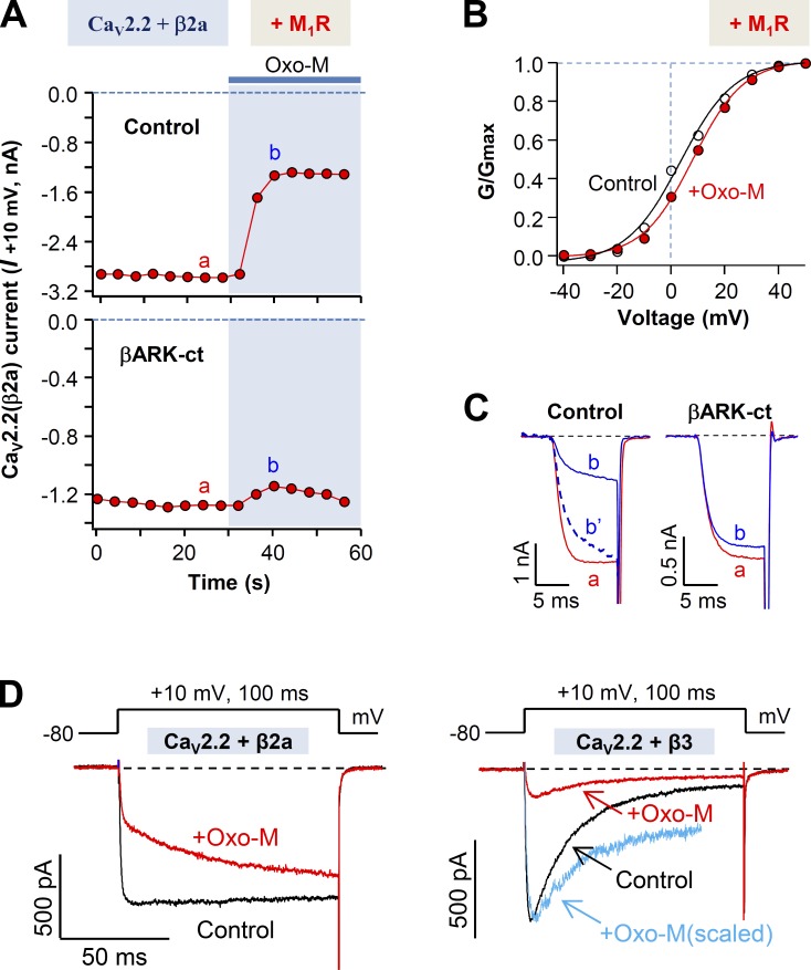
Figure 2.
βARK-ct attenuates M1 muscarinic receptor–induced inhibition of CaV2.2(β2a) currents. (A) Cells transfected with CaV2.2, α2δ1, and β2a in the presence and absence of βARK-ct were stimulated with Oxo-M, and the CaV2.2(β2a) current suppression was measured. (B) Voltage dependence of activation of the CaV2.2(β2a) channel before and during M1 receptor stimulation with Oxo-M. Dashed line is the I-V relation during Oxo-M application, which is scaled to the peak amplitude of the control. (C) Superimposed CaV2.2(β2a) current traces a and b from A. In control, the b′ dashed trace is a scaled version of b. (D) Superimposed CaV2.2(β2a) and CaV2.2(β3) current traces for control and during the stimulation of M1 receptor. Blue line in right panel shows the scaled trace of CaV2.2(β3) current after Oxo-M application (red). Note that during the Oxo-M application, activation of CaV2.2(β2a) channels (left) but not CaV2.2(β3) channels (right) is dramatically slowed.

