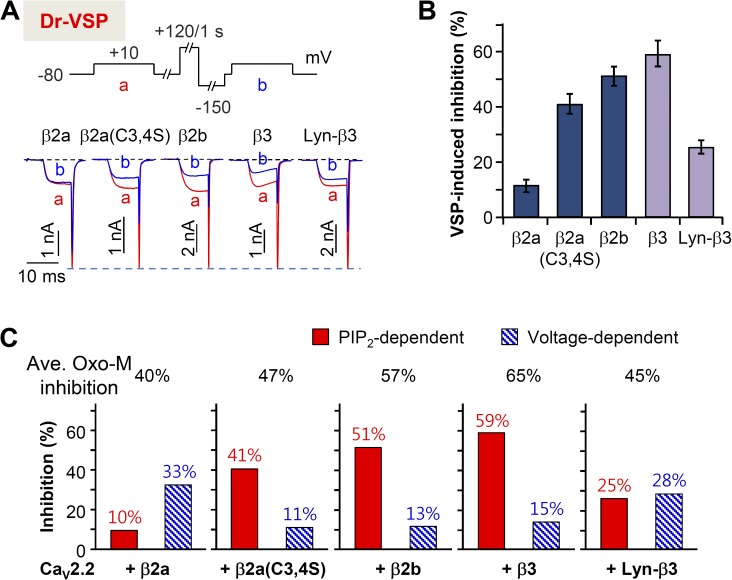
Figure 8.
Cytosolic β subunit increases the PIP2 depletion–mediated suppression of CaV2.2 currents. (A) Current inhibition by Dr-VSP activation in cells expressing different β subunits. Cells received a test pulse (a) and then were depolarized to 120 mV for 1 s, followed by the second test pulse (b). The a and b currents are superimposed. (B) Summary of the Dr-VSP–induced inhibition of CaV2.2 current. Data are mean ± SEM (n = 6–8). (C) Differential effects (mean percent inhibition) of Dr-VSP-induced, voltage-independent (A) and Gβγ-mediated, voltage-dependent (Fig. 7 B) pathways on the Oxo-M suppression of CaV2.2 channels with different CaV β subunits. Mean maximal inhibition by M1 receptor activation is presented in the top.

