Figure 1.
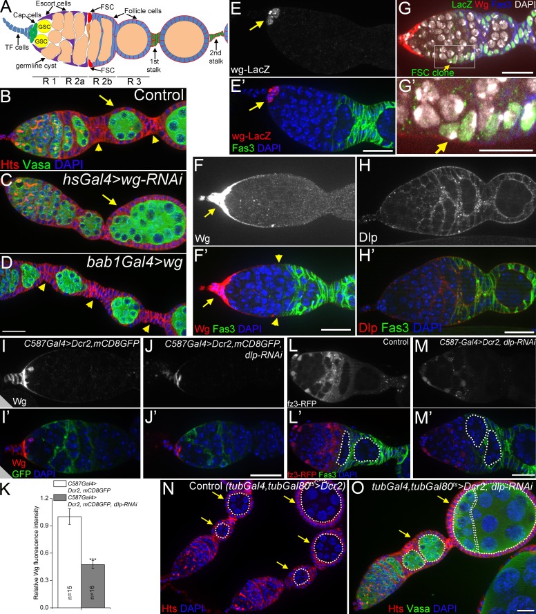
Dlp promotes long-range Wg signaling to FSCs. (A) A schematic diagram of the germarium. FSCs reside at the border of regions 2a (R 2a) and 2b. A cross-migrating FSC daughter is shown in orange. Follicle precursor cells are located in R 2b. TF cells (blue) and cap cells (green) are collectively called apical cells. GSC, germline stem cell. (B–D) Loss-of-function of wg (C) caused fused egg chambers (compound follicles, arrows; 60% penetration was observed in 35 ovarioles), whereas wg overexpression (D) resulted in stalks with increased cell numbers (arrowheads). (E) In wild-type germaria, wg was expressed only in cap cells (arrows) as shown by the wg-lacZ enhancer trap line. 3–5 cells were stained in 39/47 germaria. (F) In wild-type germaria, a continuous path of extracellular Wg (arrows), visualized at high exposure, spread to the FSCs (arrowheads). (G and G′) Wg spreading to a lacZ-labeled FSC clone (arrows). (H) Anti-Dlp in wild-type germaria. (I–K) Loss of function of dlp (with RNAi) reduced the levels of anti-Wg extracellular staining. mCD8GFP shows the pattern of C587Gal4 expression (strong in escort cells and weak in follicle cell precursors in region 2b). Dicer-2 (Dcr2) enhanced RNAi efficiency. ***, P < 0.001 (Student’s t test). Error bars represent SEM. n = number of germaria imaged. (L–M) dlp RNAi in escort cells decreased activity of the Wg signaling reporter fz3-RFP in posterior escort cells and FSCs, and caused encapsulation defects (58.3% of 84 germaria examined). 16-cell germline cysts in region 2b and region 3 are outlined. Note the two side-by-side cysts in dlp RNAi indicating an encapsulation defect. (N and O) Ubiquitous knockdown of dlp with tubGal4 resulted in fused egg chambers in 22.1% of 68 dlp RNAi germaria (arrows in O), compared with 0% in 42 control germaria (N). 16-cell germline cysts are outlined, egg chambers are indicated by arrows. DAPI labels DNA in blue. Anti-Hts labels follicle cell plasma membranes, spectrosomes, and fusomes. Anti-Vasa labels the germline. Anti-Fas3 labels follicle cell borders. Bars: (G′) 10 µm; (all other panels) 20 µm.

