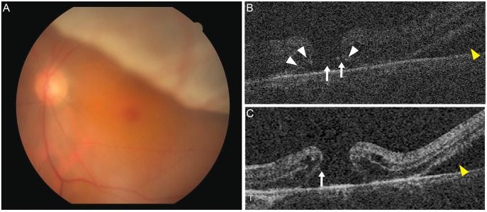
Fig. 4.
Fundus photograph and optical coherence tomography obtained from patient 4. (A) Fundus photograph showed fovea-off rhegmatogenous retinal detachment. (B) Postoperative day 8. Retinal cleavage (arrowhead) and outer retina tissue or precipitate-like materials (arrow) were detected with residual fluids (yellow arrowhead). (C) Postoperative day 15. Retinal tissues diminished further at the fovea, forming a full-thickness macular hole with perifoveal edema. Precipitate was seen on the lateral wall of hole (arrow). Subretinal fluid (yellow arrowhead) was remained.

