
Fig. 2.
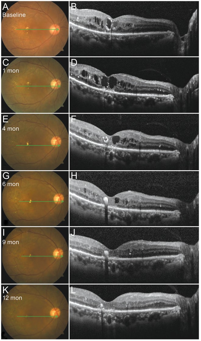
Color fundus photographs and spectral domain optical coherence tomography images of the macula for case 1. The right eye of a 63-year-old woman before (A,B) and after subthreshold micropulse yellow laser photocoagulation (C-L).
Official websites use .gov
A
.gov website belongs to an official
government organization in the United States.
Secure .gov websites use HTTPS
A lock (
) or https:// means you've safely
connected to the .gov website. Share sensitive
information only on official, secure websites.
Color fundus photographs and spectral domain optical coherence tomography images of the macula for case 1. The right eye of a 63-year-old woman before (A,B) and after subthreshold micropulse yellow laser photocoagulation (C-L).