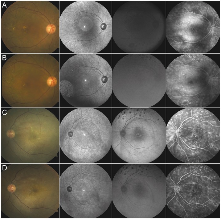
Fig. 4.
Baseline fundus color photographs, red-free photographs, autofluorescence (AF) and fluorescein angiography (FA) images for (A) case 1; (B) shows images of the same patient 14 months after subthreshold micropulse yellow laser photocoagulation (SMYLP; note the invisible chorioretinal scar in the color photograph, red-free photograph, and AF images, and the reduced fluorescein leakage in the FA image); (C) shows case 2 before treatment and (D) shows images of the same patient six months after SMYLP treatment (note that there is no chorioretinal damage in any of the images and an improved petaloid pattern of hyperfluorescence is observed in the AF and FA images).

