Fig. 2.
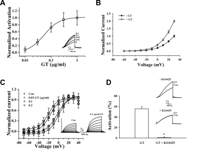
Effects of gintonin on IKs channel activity. (A) Gintonin concentration-response curves for IKs channels (mean ± S.E.M; n = 7–10 oocytes each). Inset, representative traces of gintonin-mediated IKs (KCNQ1 + KCNE1) channel activation at various gintonin concentrations. The representative peak outward current amplitude at +30 mV from a holding potential of −80 mV was measured in the absence or presence of gintonin. (B) Effects of gintonin (0.1 and 0.3 μg/ml each) on the current–voltage (I–V) relationship of the IKs channels (mean ± S.E.M; n = 10–12 oocytes each). (C) Voltage-dependence activation curves for the IKs channel. Inset, Left and right current traces are before and after application of 0.1 μg/mlgintoninto IKschannels, respectively. Currents recorded during 3 s depolarizing pulses to membrane potentials of −60 to +50 mV, applied from a holding potential of −80 mV. Tail currents were measured at −70 mV. I–V relationships for normalized IKs tail currents. Data were fitted to a Boltzmann function. (D) Attenuation of gintonin-induced IKs channel activity after treatment with Ki16425. The histogram shows blockage of gintonin-mediated IKs channel activation by the LPA1/3 receptor antagonist, Ki16425. Application of 0.1 and 1 μg/ml gintoninto IKs channels, respectively (mean ± S.E.M; n = 10–12 each) (*P < 0.001, compared to gintonin treatment only). Inset, Currents traces recorded in the absence and presence of 1 μM Ki16425 in oocytes expressing IKs channels; currents were recorded with a 3-s voltage step to +30 mV from a holding potential of −80 mV.

