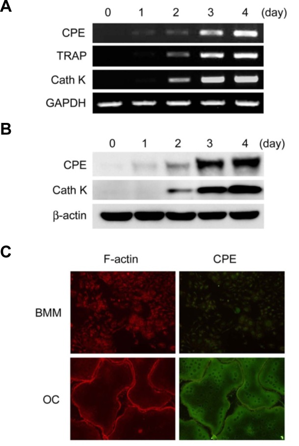
Fig. 1.

Expression of CPE is enhanced during osteoclast differentiation. BMMs were cultured in M-CSF (10 ng/ml) and RANKL (20 ng/ml) for the indicated number of days. (A) Expression of CPE was determined by RT-PCR. (B) CPE expression was analyzed by immunoblotting. TRAP or cathepsinK (CathK) served as positive controls for osteoclastogenesis, and GAPDH or β-actin served as loading controls. (C) BMMs or osteoclasts (OC) were fixed and permeabilized prior to staining. F-actin and CPE were stained with TRITC-conjugated phalloidin (red) and anti-CPE antibody (green), respectively.
