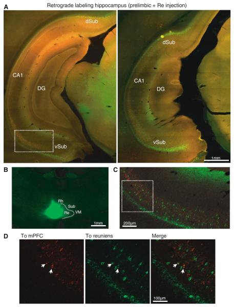
Fig. 6.
Subicular populations projecting to reuniens and mPFC. a Retrograde labeling in caudal hippocampus after CTB-488 injection in reuniens and CTB-594 injection in the prelimbic cortex (coronal sections, 5×). b Example of CTB injection in the nucleus reuniens (2.5×). c Confocal micrograph of the boxed area in (a) showing the mPFC (red) and reuniens (green) populations. d Close-up view of the boxed area in (c) with examples of double-labeled cells indicated by arrows. DG dentate gyrus; dSub dorsal subiculum; vSub ventral subiculum; Rh rhomboid; Re reuniens; Sub submedial; VM ventral medial

